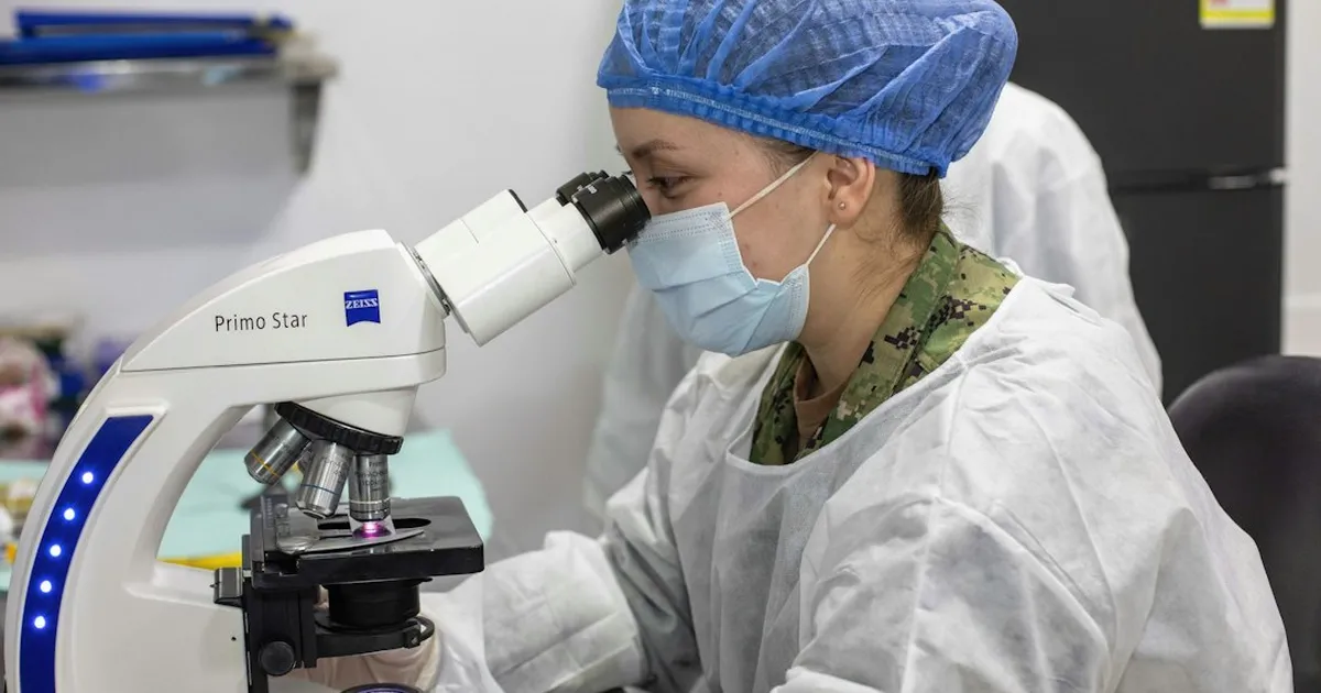
The study published in Light: Science & Applications is by Michelle Küppers, a postdoctoral researcher in W. E. Moerner's laboratory, and Moerner, a professor of chemistry and also of applied physics at Stanford University. The technique is called interferometric image scanning microscopy, or iISM. It combines interferometric scattering microscopy (iSCAT), which detects nanoscale structures through their light scattering, with image scanning microscopy (ISM) to improve spatial resolution.
According to Küppers and Moerner, conventional fluorescence imaging can suffer from phototoxicity, limited labeling efficiency or disruption of biological functions. iSCAT avoids dyes and genetic markers but encounters speckle in living cells, arising from the coherent superposition of scattering events in complex, heterogeneous media.
How the microscope works without dyes
According to an account by the Stanford School of Humanities and Sciences, in interferometric scattering microscopy, a laser illuminates the cell, whose small structures scatter some of the light, while a second laser beam amplifies the weaker scattered light until it can be detected. According to a report by Scientist Live, iISM replaces the single confocal pinhole detector with an array detector that records the full interferometric point spread function at each scan position and captures many off-axis pinholes in parallel. To accommodate this point spread function, Küppers and Moerner developed their own adaptive pixel reassignment algorithm, which combines the parallel measurements into a reconstructed image with an improved contrast-to-noise ratio. To avoid a point spread function that is often anisotropic under linearly polarized light, the authors also used circularly polarized light, which, according to a review article by Liang, Ren and Xi, ensures the rotational symmetry of the interferometric point spread function and reduces measurement artifacts.
What the measurements show
For calibration, the researchers used 60-nanometer polystyrene nanoparticles distributed on a coverslip. With iISM and the modified algorithm, they achieved a lateral resolution limit of 120 nanometers ± 4 nanometers at a noise level of about 0.008. In the same setup, at the same incident illumination power, iISM achieved a contrast-to-noise ratio of about 38, almost four times higher than with a closed confocal pinhole and about three times higher than with an open one. According to the authors, the incident illumination power required per diffraction-limited light spot is around 10 times lower than in conventional confocal microscopy. The researchers say they can use iISM to visualize the endoplasmic reticulum, actin cytoskeleton, mitochondria and vesicles in living cells with what they describe as practically unlimited observation times; their correlative images of the actin cytoskeleton using fluorescence ISM, however, were obtained from fixed, nonliving cells. “By parallelizing interferometric imaging and using modified computational reassignment, iISM recovers information that would otherwise be discarded, allowing contrast and resolution to be improved without increasing the light dose,” Küppers said, according to Scientist Live.

In comparison: the predecessor technique
Back in 2023, Küppers and other researchers presented an earlier technique using confocally operated iSCAT in Nature Communications, for which Abbe's formula gave a theoretical resolution limit of 152 nanometers at a wavelength of 445 nanometers. Using this technique on a HeLa cell, they localized the basal nuclear envelope laterally with an accuracy of 180 nanometers. Two artificial chromium nanopillars spaced 130 nanometers apart could be clearly resolved at a narrow pinhole setting of 0.3 AU, while a setting of 1.2 AU had difficulty doing so. A roadmap for label-free super-resolution imaging published in October 2023 describes the broader research field of label-free super-resolution. The authors of the iISM study themselves acknowledge that a complete theoretical description of the interferometric point spread function is beyond the scope of their work. A report by ScienceDaily states that it is based on material provided by Stanford University. According to SciTechDaily, iISM also does not achieve the resolution of some highly specialized microscopes; first author Küppers advocates using it alongside fluorescence microscopy: fluorescence provides molecular specificity, while iISM provides label-free context and observation of dynamics.
Moerner's next goal, according to an April 2, 2026 report: faster imaging
“Our next goal is to develop iISM further toward faster dynamics and enable widespread adoption through higher imaging speed and broader accessibility,” Moerner said, according to Scientist Live. According to a ScienceDaily report dated October 16, 2026, Moerner and Küppers are working to further develop the instrument, with the aim of making it accessible to researchers from many areas of biology in the future. According to SciTechDaily, as of its May 31, 2026 report, three collaborations with other Stanford research groups were already underway: one on interactions between plant cells, fungi and bacteria, one on the entry of a cancer drug into a cell, and a project still in the planning stage on changes in the shape of red blood cells during a malaria infection. According to Stanford University, the research was supported by the U.S. National Institute of General Medical Sciences.